Soft X-ray microscopy is a powerful tool for characterization of various material properties in a large range of applications. It can provide insights into the nanostructure of thin organic films for solar cell or battery applications, but also reveal complex spin dynamics in novel nanomagnetic systems for future data memory technologies. In-situ imaging of chemical processes in cells, catalytic reactions on particles relevant for atmospheric chemistry and imaging of geological specimens are amongst other typically investigated specimens. In many cases, nanostructures in the size range of 10 – 50 nm play an important role, requiring significantly better resolution for an optimum microscopy technique.
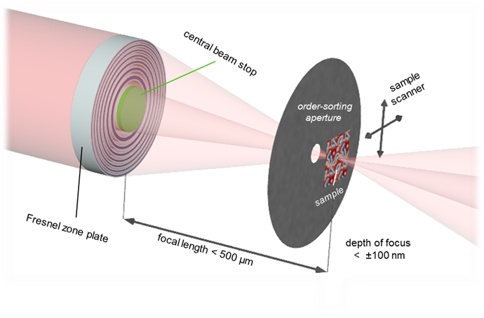
The short wavelength of soft X-rays provides a potential resolution limit of 1 – 5 nm according to the Abbe diffraction limit. However, diffraction limited imaging has so far not been achieved in soft X-ray imaging despite considerable efforts. The barrier of 10 nm has only been surpassed by indirect imaging techniques requiring image reconstruction, while standard experiments usually work with a resolution of more than 25 nm. The resolution limit in direct soft X-ray imaging is mainly pushed by improvements in the design and lithography-based fabrication procedure of the required diffractive focusing optics, so-called Fresnel zone plates.
An international team of researchers from the Paul-Scherrer-Institute in Villigen, Switzerland (Dr. Christian David, Dr. Jörg Raabe and FAU-Alumnus Dr. Benedikt Rösner), the Chair of Physical Chemistry II at FAU (Prof. Dr. Rainer Fink and Dr. Andreas Späth), and further institutes in Paris, Hamburg and Basel report a record resolution of 7 nm for soft X-ray direct imaging. Sub-10 nm resolution has been achieved in several experiments at the soft X-ray microscopes PolLux (Swiss Light Source, Switzerland) and HERMES (Synchrotron SOLEIL, France). A remarkable feature of this study is that record resolution was not only achieved with specifically designed prototype specimens, but also real scientific challenges could be addressed by real-space imaging of magnetic coupling in agglomerates of iron nanoparticles with a size distribution of 5 – 20 nm. The study has been published in Optica, the flagship journal of the Optical Society of America (DOI: 10.1364/OPTICA.399885).
The collaborators expect this significant improvement of resolution in soft X-ray microscopy to trigger a large number of cutting-edge studies especially in solar cell and nanomagnetic applications, since the single-digit nanometer range is highly relevant in those fields.
The project received funding by the German Federal Minister of Education and Research (BMBF, grants 05K16WED and 05K19WE2), DFG grant SP 1775/1-1 and the EU-H2020 Research and Innovation Program.
Contact
Prof. Dr. Rainer Fink
rainer.fink@fau.de